YOUR CART
- No products in the cart.
Subtotal:
₹0.00
Anand Eye Institute is a tertiary care eye hospital, a teaching institute for medical, surgical, para-medical and non-clinical or administrative skills, a socially responsible company (through its charitable SAVERANAND NETHRALAYA) and a place for healing and tranquillity; Specialist surgeons, trained and practised in individual specialisations, Latest equipment, supporting infrastructure and facilities bundled into a 5 storey, 50 bedded, 16000 SFT lavish structure.

The new Angio OCT is designed to prepare an angiogram report without the use of the Fluroscein dye. OCT-Angiography (OCTA) – is a new way of seeing the presence of blood flow in the vessels of the eye. It enables ophthalmologists to assess ocular diseases with unprecedented detail The AngioVue is the only commercially available OCTA system capable of imaging the structure of the small blood vessels of the eye, through a non-invasive procedure. Meaning, the revolutionary new machine takes high quality 3d images without the need of the dye injection. OCT is a well-established medical imaging and diagnostics technique used to understand the condition of the different layers of the Retina, Choroid and Cornea which are multi layered structures. OCT projects a 3D image of all the layers, with the help of which ophthalmologists can arrive at a diagnosis and plan further treatment.
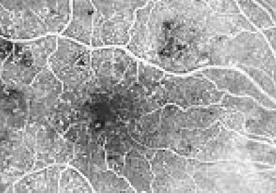
Angiography is a medical imaging and diagnostics technique for visualizing the blood vessels. FFA is a similar procedure for visualizing the vessels in the retina and the choroid. A small amount of yellow – fluorescein dye is injected into the arm that travels to the vessels of the eye, which when illuminated with blue light shows clearly demarcated vessels, which are photographed by specialised camera. Most commonly advised for Vitreous haemorrhage, Age related Macular Degeneration and advanced diabetic retinopathy.
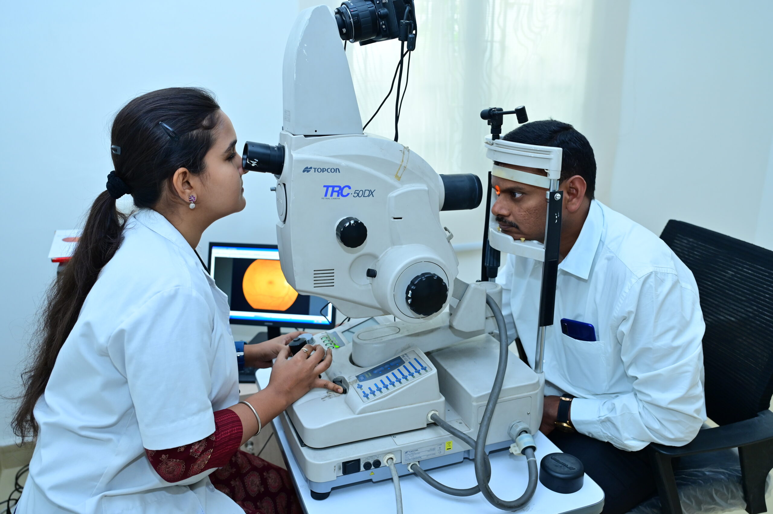
The Fundus of the eye is the entire inside surface of the eye comprising of Retina, Macula, Optic disc and Posterior pole. Photographs are taken by a high definition Digital SLR camera mounted on an intricate arrangement of reflective mirrors that take accurate pictures from different angles. Fundus photos in combination with Fundus Angiography is used for diagnosis of Glaucoma and various retinal disorders of Retina.

Corneal topography is a special photography technique that maps the surface of the clear, front window of the eye (the cornea). It works much like a 3D (three-dimensional) map. With a topography scan, a doctor can find distortions in the curvature of the cornea, which is normally smooth. It also helps doctors monitor eye disease and plan for LASIK/ REFRACTIVE surgery.
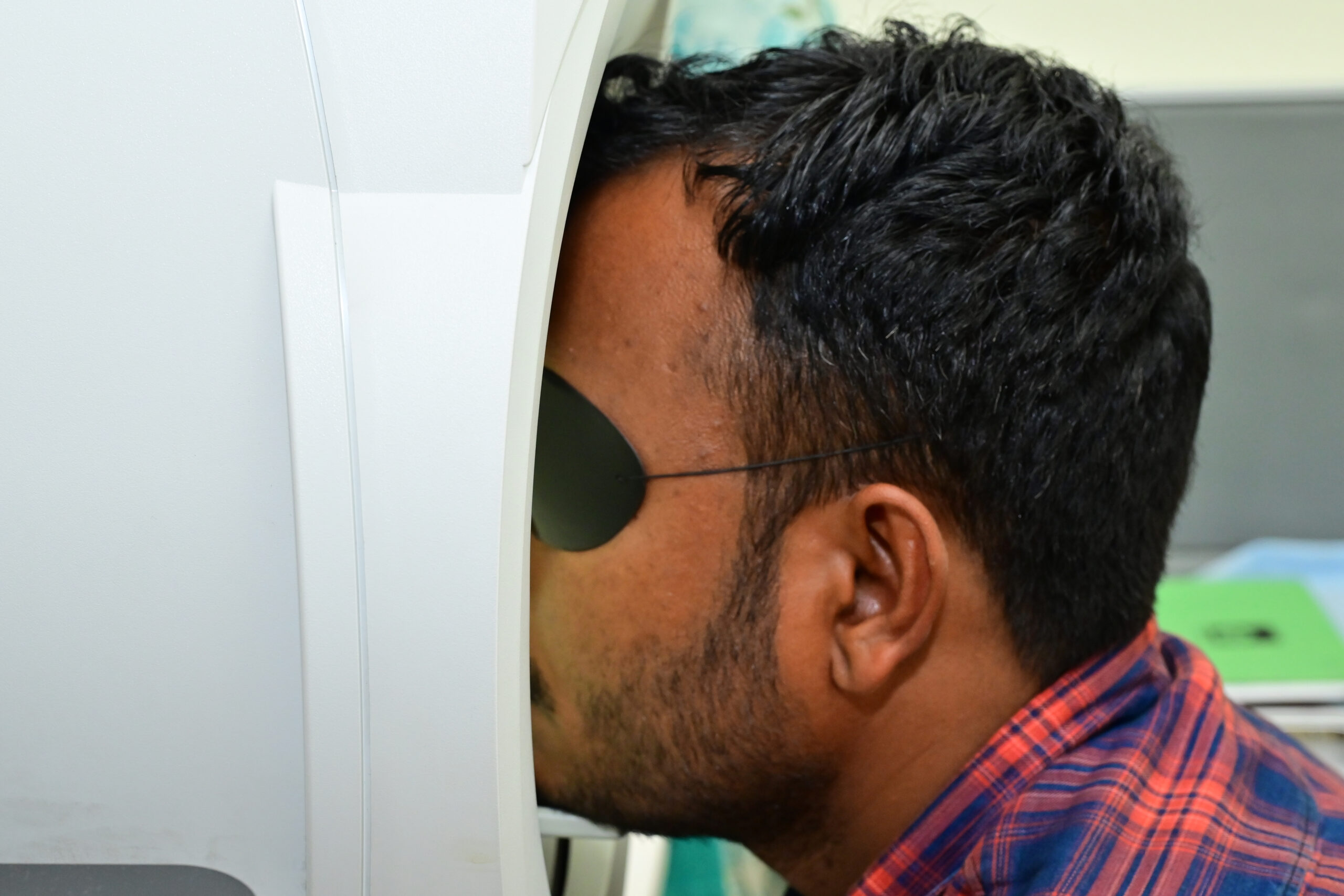
It is a tool for measuring the human visual field that is commonly used. It is also called as Automated Perimetry. The results of the analyzer identify the presence of any defects in vision defect. This guides the doctors is diagnosing as well monitoring progression of optic nerve related diseases like Glaucoma etc.
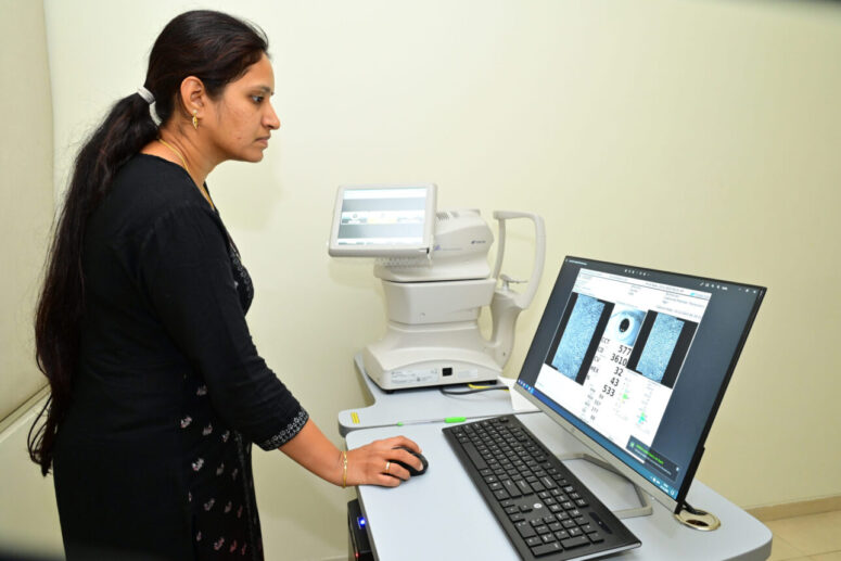
Specular microscopy is a non-invasive modality to document the healthy and diseased endothelium photographically. Specular microscopy is also crucial in assessing the preoperative endothelial health before high-risk surgeries, comparing various techniques, the impact of lasers during refractive surgery, and the assessment of donor cornea before transplantation.

This is ultrasonography of the eye. High frequency sound waves are used to look inside the eye. This is similar to ultrasound performed for other parts of the body. This test is particularly helpful when the doctor has a doubt and is not able to visualise the inner structures during a normal examination.
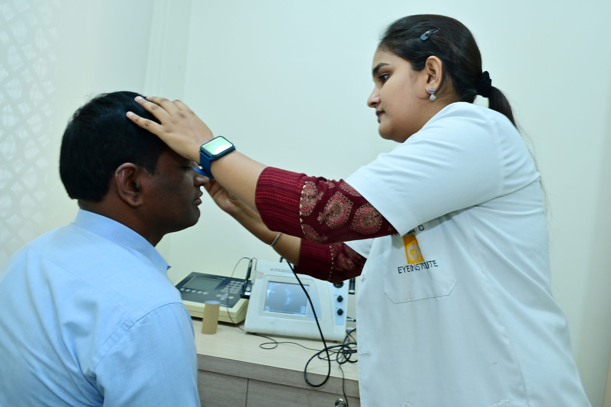
Ascan biometry also uses ultra sound waves to determine the axial length of the lens in cataract for calculating the power of IOL that has to be used in place of the Cataractous lens.
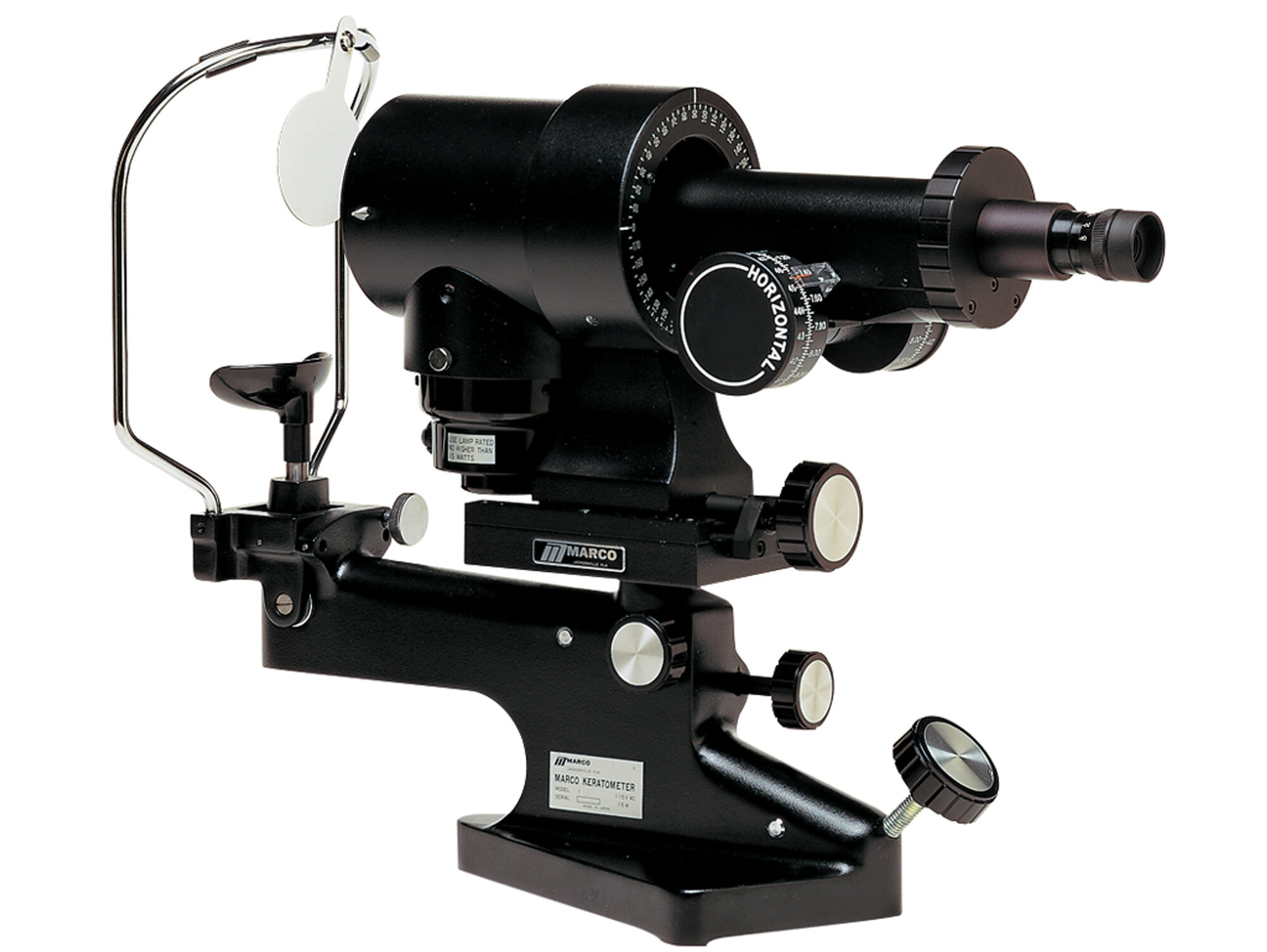
This is used to measure the curvature of the cornea, that indirectly determines the refractive power of the Lens to be used.